Overview
- Eyes have evolved independently dozens of times across the animal kingdom, yet all animal eyes share a common molecular foundation: the Pax6 master control gene and opsin photopigments, indicating descent from a single ancestral light-sensing patch.
- A continuous spectrum of functional eye types exists in living organisms today, from simple light-sensitive eyespots in single-celled organisms through pit eyes and pinhole cameras to the sophisticated lens eyes of vertebrates and cephalopods, each stage conferring a selective advantage.
- Computational modeling shows that a camera-type eye can evolve from a flat photosensitive patch in fewer than 400,000 generations under conservative assumptions, and the fossil record confirms that complex compound eyes were already present by the early Cambrian, over 500 million years ago.
The eye is among the most frequently cited examples of biological complexity, and its evolution has been studied in greater detail than perhaps any other organ system. Far from presenting an insurmountable challenge to evolutionary theory, the eye has become one of its strongest case studies. A wealth of comparative anatomy, molecular genetics, computational modeling, and fossil evidence now documents how complex image-forming eyes evolved through a long series of intermediate stages, each conferring a selective advantage on its possessor.7, 8
Living organisms today display a remarkably complete spectrum of eye types, from the simplest light-sensitive patches of single-celled protists to the sophisticated camera eyes of vertebrates and cephalopods. Molecular studies have revealed that this diversity rests on a shared genetic foundation: the same master regulatory gene, Pax6, controls eye development across virtually all animal phyla, and the light-detecting opsin proteins found in every animal eye descend from a common ancestor that predates eyes themselves.6, 9
Darwin and the eye
Charles Darwin addressed the evolution of the eye directly in On the Origin of Species (1859), in a chapter titled "Difficulties on Theory." He wrote that to suppose the eye "could have been formed by natural selection, seems, I freely confess, absurd in the highest possible degree." This sentence, frequently quoted out of context, was in fact the opening of an extended argument for the eye's evolution by natural selection. Darwin immediately continued: "Yet reason tells me, that if numerous gradations from a perfect and complex eye to one very imperfect and simple, each grade being useful to its possessor, can be shown to exist... then the difficulty of believing that a perfect and complex eye could be formed by natural selection, though insuperable by our imagination, can hardly be considered real."1
Darwin then catalogued the diversity of eyes in living animals, from aggregates of pigment cells with no nerve at all, to simple optic nerves coated with pigment, to more elaborate structures with refractive bodies. He argued that these represented a plausible series of intermediate stages, each functional and each conferring a selective advantage over the previous form. The key insight was that an eye need not be "perfect" to be useful: even a crude ability to detect light and shadow provides information about predators, prey, and the environment that an eyeless organism lacks entirely.1
Darwin's reasoning was prescient but necessarily limited by the knowledge available to him. He could not have known about the molecular genetics of eye development or the Cambrian fossil record of early eyes. Modern research has not only confirmed his core argument but has filled in the intermediate stages with far greater precision than he imagined possible.16
The spectrum of eye types in living organisms
One of the most compelling lines of evidence for eye evolution comes from the diversity of eye types found in living animals today. These range from the simplest conceivable light-sensing structures to highly sophisticated image-forming organs, and every intermediate grade is represented by at least one living species. This does not mean that modern organisms are ancestral to one another; rather, each represents an independent solution to the problem of extracting spatial information from light, preserved because each solution is adaptive in its own ecological context.8, 15
At the simplest level, many single-celled organisms possess eyespots: small patches of photosensitive pigment that allow the cell to detect light direction. The euglenid Euglena, for example, has a stigma (a carotenoid-containing granule) adjacent to the base of its flagellum that functions as a directional light sensor, enabling the organism to swim toward or away from light sources. No lens, no image, no nerve cell is required. The eyespot is simply a patch of pigment that shades a photoreceptor from one side, creating a directional signal.8
Among multicellular animals, flatworms (planarians) possess cup-shaped pigment eyes. These consist of a small cluster of photoreceptor cells set into a cup of dark pigment. The cup shape means that light from different directions strikes different photoreceptor cells, giving the animal crude directional vision. Planarians use these eyes primarily to avoid light, retreating under rocks and into crevices. The pigment cup eye represents a modest but significant advance over a flat eyespot: it provides spatial information about where light is coming from, not merely whether light is present.8, 15
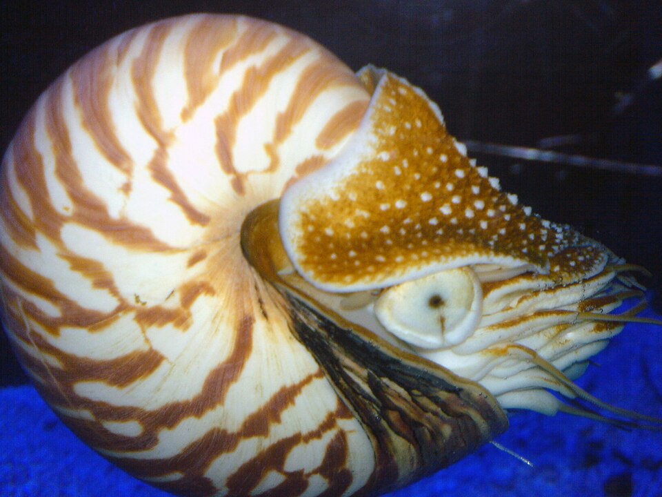
A deeper cup creates a pinhole eye, exemplified by the chambered nautilus (Nautilus pompilius). The nautilus eye is essentially a deep pit open to the sea, with no lens. Light enters through a small aperture and projects an image (albeit dim and blurry) onto a retina lining the back of the cup. The pinhole camera principle means that a smaller aperture produces a sharper image but admits less light, creating a fundamental trade-off. The nautilus eye is fully functional for the animal's deep-water, low-light lifestyle, but it cannot match the resolving power of a lens eye.8
The addition of a lens dramatically improves both resolution and light-gathering power. Lens eyes are found in vertebrates, cephalopods (squids and octopuses), some snails, and certain spiders. In each case, a transparent refractive structure focuses light onto a layer of photoreceptor cells, forming a sharp image. The lens can be a simple sphere (as in many fish) or a more complex aspherical structure with a graded refractive index (as in the human eye). Compound eyes, found in arthropods such as insects and crustaceans, achieve wide-angle vision through an array of many small optical units (ommatidia), each sampling a different direction.7, 8
Functional spectrum of eye types across living organisms8, 15
Opsins: light-sensitive proteins that predate eyes
The molecular basis of all animal vision is the opsin protein family. Opsins are membrane-spanning proteins that bind a light-absorbing chromophore (typically retinal, a derivative of vitamin A) and undergo a conformational change when struck by a photon, initiating a signaling cascade within the photoreceptor cell. All known animal opsins share a common ancestor, and molecular phylogenies reveal that the major opsin families diverged before the evolution of complex eyes.6, 17
Three broad classes of opsin are recognized in animals: ciliary opsins (c-opsins), rhabdomeric opsins (r-opsins), and a group sometimes called RGR/Go opsins. Phylogenetic analyses indicate that all three classes were already present in the last common ancestor of bilaterally symmetrical animals (Bilateria), which lived roughly 600 million years ago, well before the Cambrian explosion that produced the first complex eyes in the fossil record.6, 14
Crucially, opsins did not originally evolve for vision. In many organisms, opsins play roles in circadian rhythm regulation, phototaxis (movement toward or away from light), and other non-visual light responses. Cnidarians such as jellyfish and corals possess opsins and simple photoreceptors but lack anything resembling an image-forming eye. The cnidarian opsin system appears to function primarily in regulating spawning cycles and diel vertical migration in response to ambient light levels.13, 17 This pattern is consistent with the evolutionary principle of co-option (also called exaptation): a molecular system that initially evolved for one function was subsequently recruited for a different, more complex function when combined with new anatomical structures.7
The deep antiquity of opsins means that the molecular machinery for detecting light was already in place long before natural selection began shaping it into the elaborate optical systems seen in modern animals. Eyes did not need to invent photoreception from scratch; they built upon a pre-existing molecular toolkit.6, 9
Pax6: the master control gene for eye development
Perhaps the most striking molecular evidence for the common origin of animal eyes is the transcription factor Pax6. In 1994, Quiring and colleagues demonstrated that the eyeless gene of the fruit fly Drosophila melanogaster is homologous to the Small eye gene in mice and the Aniridia gene in humans. All three are versions of the same gene, Pax6, and mutations in any of them produce analogous eye defects: reduced or absent eyes in flies, small eyes in mice, and absent irises in humans.3
The following year, Halder, Callaerts, and Gehring performed a landmark experiment. They used genetic techniques to express the eyeless gene (the fly version of Pax6) in tissues where it is not normally active, such as the legs, wings, and antennae of Drosophila. The result was the formation of ectopic (misplaced) compound eyes on these body parts. Remarkably, when the mouse version of Pax6 was expressed in fly tissues, it too induced the formation of ectopic compound eyes, not mouse eyes, demonstrating that the gene's role as a master switch for eye development is conserved across more than 500 million years of independent evolution.4
Pax6 functions as a master regulatory gene: it sits at the top of a developmental cascade that activates roughly 2,500 downstream genes required for eye morphogenesis.12 Subsequent research has found Pax6 homologs controlling eye development in organisms as diverse as squids, sea urchins, ribbonworms, and planarian flatworms. The universality of Pax6 strongly suggests that the last common ancestor of all bilaterian animals already possessed a Pax6-dependent light-sensing system, a proto-eye, from which the diverse array of modern eye types descended.9, 12
This finding resolves an apparent paradox. If eyes evolved independently dozens of times, as comparative anatomy suggests, how can they all share the same master control gene? The answer is that what evolved independently was not the genetic toolkit for light sensing (which is ancient and shared) but the downstream elaboration of that toolkit into different optical designs: compound eyes, camera eyes, mirror eyes, and others. The independent origins are real, but they were all built on the same molecular foundation.7, 12
Computational modeling of eye evolution
In 1994, the same year that Pax6 was discovered in flies, the Swedish biologists Dan-Eric Nilsson and Susanne Pelger published a computational model asking how long it would take for a camera-type eye to evolve from a flat patch of light-sensitive cells. Their paper, titled "A pessimistic estimate of the time required for an eye to evolve," deliberately adopted conservative assumptions at every step to produce an upper bound on the time required.2
The model began with a flat layer of photoreceptor cells sandwiched between a transparent protective layer above and a pigmented layer below. Through a series of small, quantifiable morphological changes, each improving the optical performance of the structure by at least one percent, the model traced the transformation from flat patch to a cup eye, then to a pinhole eye, and finally to a lens eye with a graded refractive index. Each step involved only changes in tissue geometry (curvature, aperture size) or refractive index, all of which are known to vary within natural populations.2
Under their pessimistic assumptions, including a low coefficient of variation (0.01), a heritability of 0.50, and a selection intensity of only 0.01 per generation, Nilsson and Pelger calculated that the entire transformation from flat patch to camera eye would require approximately 364,000 generations. For a small aquatic animal with a generation time of one year, this corresponds to fewer than 400,000 years, a geological instant given that animal life has existed for over 600 million years. Even with extremely conservative parameters, the time required was orders of magnitude shorter than the time available.2
The Nilsson-Pelger model does not claim that eye evolution actually followed this precise sequence. Rather, it demonstrates that there is no temporal barrier to the evolution of complex eyes by natural selection: even under worst-case assumptions, the process is surprisingly fast. This finding is consistent with the observation that complex eyes appear abruptly in the early Cambrian fossil record, as the "light switch" hypothesis proposes, suggesting that once the ecological conditions favored vision, eyes evolved rapidly.2, 18
Convergent evolution of eyes
Eyes have evolved independently more times than almost any other complex organ. In a landmark 1977 survey, Salvini-Plawen and Mayr catalogued eye types across the animal kingdom and concluded that photoreceptors and eyes had arisen independently at least 40, and possibly as many as 65, separate times.5 More recent analyses, incorporating molecular data, have generally supported estimates in this range, though the exact number depends on how "eye" is defined and whether one counts only image-forming eyes or also simpler photoreceptive organs.7, 15
The most celebrated example of convergent evolution in eyes is the camera eye, which evolved independently in vertebrates and in cephalopod mollusks (octopuses and squids). Both possess a single-chambered eye with a cornea, an adjustable iris, a crystalline lens, and a retina, yet these structures evolved from entirely different tissue types in each lineage. Vertebrate lenses are composed of crystallin proteins recruited from heat-shock proteins and metabolic enzymes, while cephalopod lenses use a different set of proteins. The optical result is strikingly similar, but the underlying molecular components differ, exactly as convergent evolution predicts.7, 8
Compound eyes, the dominant visual system in arthropods, also evolved independently from camera eyes. The insect compound eye consists of hundreds to thousands of ommatidia, each containing its own lens and a small cluster of photoreceptor cells. This design excels at detecting motion and provides an extremely wide field of view, but it sacrifices the spatial resolution that camera eyes achieve. Some arthropod lineages, such as the mantis shrimp, have evolved compound eyes of extraordinary complexity, with 16 types of photoreceptor cells compared to the three or four found in most vertebrates.8
The repeated, independent evolution of eyes is not surprising given the universal utility of vision. Light is the most information-rich environmental signal available, and any organism that can extract spatial information from light gains an immediate selective advantage for finding food, avoiding predators, and navigating its environment. The physics of light constrains the possible optical solutions to a small number of workable designs, which is why the same basic eye types (camera, compound, mirror, scanning) appear again and again across unrelated lineages.7, 15
The vertebrate retina and its inverted wiring
The vertebrate retina has a peculiar arrangement that makes sense in light of evolutionary history but would be puzzling if eyes were designed from scratch. In the vertebrate eye, light must pass through several layers of neurons, including ganglion cells, amacrine cells, and bipolar cells, before reaching the photoreceptor cells (rods and cones) at the back of the retina. The photoreceptors point away from the incoming light, toward the pigment epithelium behind them. This "inverted" or "backwards" arrangement means that the neural wiring sits between the light and the photoreceptors, and the axons of the ganglion cells must exit the eye through a hole in the retina, creating the optic disc, a region with no photoreceptors that produces a blind spot in each eye.6, 16
The cephalopod eye, by contrast, has its photoreceptors facing the incoming light, with the neural wiring behind the retina. This "everted" arrangement means that cephalopods have no blind spot and no need for light to traverse neural tissue before reaching the photoreceptors. The cephalopod design is, in purely engineering terms, the more straightforward arrangement.8
The reason for the vertebrate retina's inverted wiring lies in its evolutionary origin. Vertebrate photoreceptors are ciliary in type (derived from modified cilia), and they evolved from cells lining the interior of the neural tube. When the optic cup forms during embryonic development, it does so by an invagination (infolding) of the developing brain, which necessarily places the photoreceptor layer on the inside, facing away from the light. This developmental pathway is a historical constraint inherited from the earliest vertebrate ancestors. The resulting arrangement works perfectly well in practice, as the neural layers are largely transparent, but it is exactly the kind of imperfect, historically contingent design that evolution produces and that engineering from scratch would not.6, 9
The fossil record of early eyes
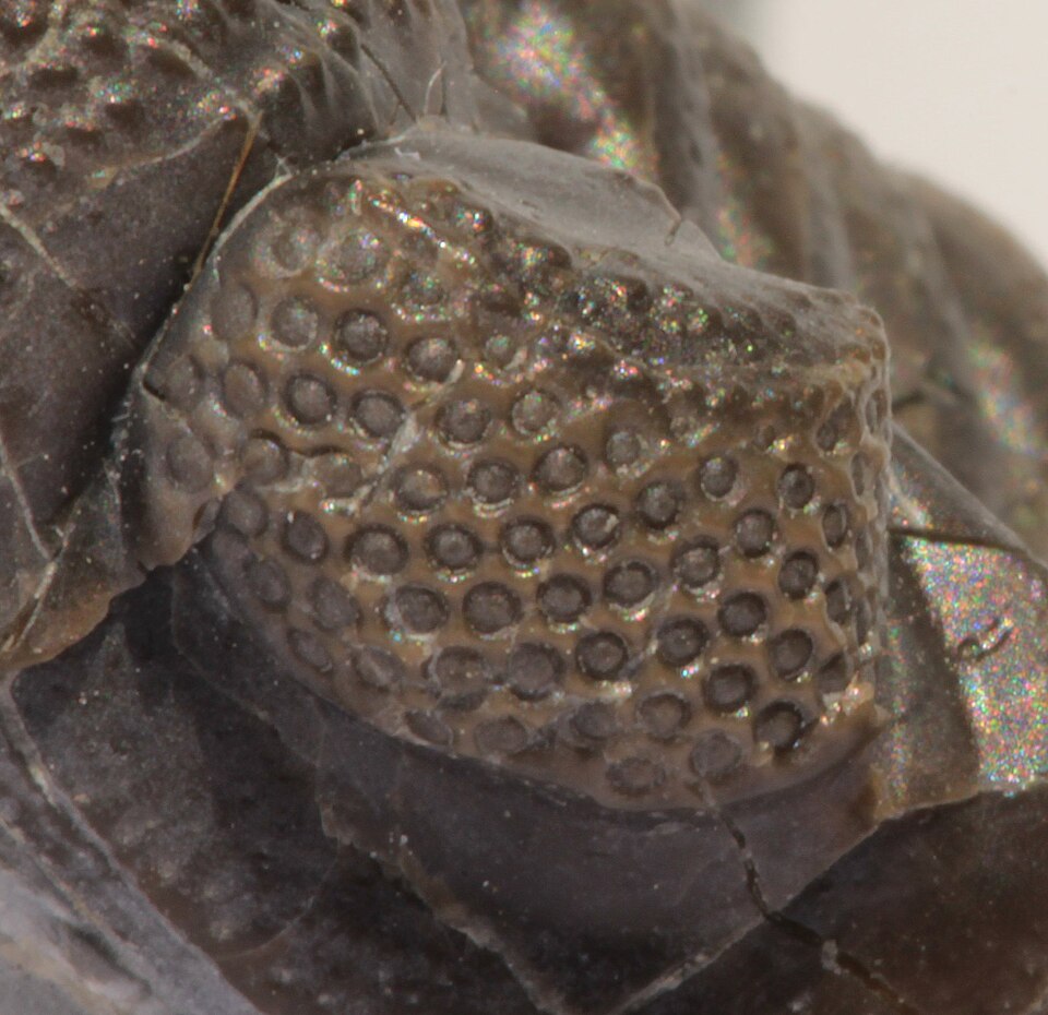
The fossil record provides direct evidence that complex eyes existed remarkably early in animal history. The oldest well-preserved compound eyes belong to trilobites, the diverse and abundant marine arthropods that dominated Cambrian seas. Trilobite eyes are unique among all known animals in that their lenses were composed of crystalline calcite (calcium carbonate) rather than organic proteins. Because calcite is a mineral, trilobite lenses fossilize readily, preserving optical details with extraordinary fidelity.10, 11
In 2017, Schoenemann, Pärnaste, and Clarkson described the internal structure of a compound eye from the lower Cambrian trilobite Schmidtiellus reetae, dated to approximately 521 million years ago. Phosphatization had preserved the cellular details of individual ommatidia, revealing a structure essentially identical to the apposition compound eyes of modern bees and dragonflies, complete with a crystalline cone and a rhabdom (the light-sensitive structure within each ommatidium). This represents the oldest known preserved visual system in the fossil record.11
Later trilobites, particularly members of the order Phacopida, evolved even more sophisticated optics. In 1975, Clarkson and Levi-Setti demonstrated that phacopid trilobite lenses contained an internal doublet structure that corrected for spherical aberration, the optical defect that causes light passing through the edges of a simple lens to focus at a different point than light passing through the center. The correction employed in these trilobite lenses follows the same mathematical principles described centuries later by Descartes and Huygens for manufactured optics.10
The sudden appearance of well-developed eyes in the early Cambrian has been linked to the broader phenomenon of the Cambrian explosion, the rapid diversification of animal body plans that occurred between roughly 540 and 520 million years ago. Andrew Parker's "light switch" hypothesis proposes that the evolution of the first image-forming eyes triggered an evolutionary arms race between predators and prey, driving the rapid diversification of body plans, hard shells, spines, and burrowing behaviors that characterize the Cambrian fossil record.18, 19
Two ancestral photoreceptor cell types
A further layer of molecular evidence comes from the discovery that animals possess two fundamentally distinct types of photoreceptor cells, each using a different class of opsin and a different signaling pathway. Ciliary photoreceptors, which expand their membrane surface area using modified cilia, are the dominant photoreceptor type in vertebrate eyes and use ciliary opsins (c-opsins) coupled to a cyclic nucleotide signaling cascade. Rhabdomeric photoreceptors, which expand their membrane surface area using microvilli (tiny finger-like projections), are the dominant type in arthropod and mollusk eyes and use rhabdomeric opsins (r-opsins) coupled to a phospholipase C signaling cascade.9, 14
Detlev Arendt and colleagues showed that both photoreceptor types are present in a single organism, the ragworm Platynereis dumerilii, a marine annelid. In this animal, rhabdomeric photoreceptors are found in the eyes, while ciliary photoreceptors are found in the brain, where they appear to function in circadian rhythm regulation rather than vision. This dual system suggests that the last common ancestor of bilaterians already possessed both photoreceptor types, one for vision and one for non-visual light sensing, which were subsequently deployed differently in different lineages.9
Vertebrates, unusually, use ciliary photoreceptors for vision and have largely lost rhabdomeric photoreceptors from their eyes, though a rhabdomeric-type opsin (melanopsin) persists in a subset of retinal ganglion cells where it mediates the pupillary light reflex and circadian photoentrainment. Arthropods and mollusks, conversely, use rhabdomeric photoreceptors for vision. This divergent deployment of an ancestral dual system explains both the deep molecular homologies between all animal eyes and the real structural differences between vertebrate and invertebrate photoreceptor cells.6, 9
Broader implications
The evolution of the eye illustrates several general principles of evolutionary biology. First, it demonstrates that natural selection can build complex organs incrementally, with each intermediate stage functional and adaptive in its own right. There is no stage in the progression from eyespot to camera eye that would be useless to its possessor; each provides information that the previous stage did not.2, 16
Second, eye evolution exemplifies the concept of deep homology: the use of shared genetic toolkits (Pax6, opsins) across distantly related lineages to build structures that are superficially very different. Deep homology blurs the traditional distinction between homologous structures (inherited from a common ancestor) and analogous structures (independently evolved), because the same underlying genes can be independently recruited to build different types of eyes in different lineages.7, 12
Third, the vertebrate retina's inverted wiring is a textbook example of historical constraint in evolution. Evolution does not design organs from a blank slate; it modifies existing structures, and the results bear the unmistakable imprint of their developmental and evolutionary history. The blind spot in the vertebrate eye is not a flaw that a designer would introduce; it is an inevitable consequence of the developmental pathway by which the vertebrate retina forms.6, 16
Finally, the speed with which complex eyes can evolve, fewer than 400,000 generations even under pessimistic assumptions, removes any intuitive objection based on the apparent improbability of such structures arising by gradual modification. The Cambrian fossil record confirms this: once ecological conditions favored vision, complex eyes appeared in multiple lineages within a geologically brief interval.2, 11, 18
References
Homology of the eyeless gene of Drosophila to the Small eye gene in mice and Aniridia in humans