Overview
- An atavism is the reappearance in an individual of an ancestral trait that disappeared from the lineage millions of years ago, caused by the reactivation of dormant developmental genes rather than the introduction of new genetic material.
- Classic examples include coccygeal projections in humans, rudimentary hind limbs in whales and dolphins, extra toes in horses, and teeth in chickens — each confirmed by fossil evidence showing the trait was once normal in the lineage.
- Atavisms are powerful evidence for common descent because they require ancestral gene networks to remain functionally intact across vast timescales, which has no coherent explanation under special creation but follows directly from descent with modification.
An atavism is the spontaneous reappearance, in an individual organism, of a trait that was present in its distant ancestors but absent for many generations. The word derives from the Latin atavus, meaning great-great-great-grandfather or, more loosely, forefather. In biology, atavisms are not mere curiosities of development. They are windows into the evolutionary past—direct evidence that the genetic instructions for building ancestral structures remain embedded in an organism’s genome long after those structures ceased to be expressed, preserved in functional form across millions of years of descent. A dolphin that is born with small, externally visible hind limb buds, a horse that develops a full three-toed foot, or a human infant born with a fleshy tail extending beyond the coccyx is not the product of a new mutation. Each is the product of an ancient developmental program reawakened by a shift in gene regulation.13
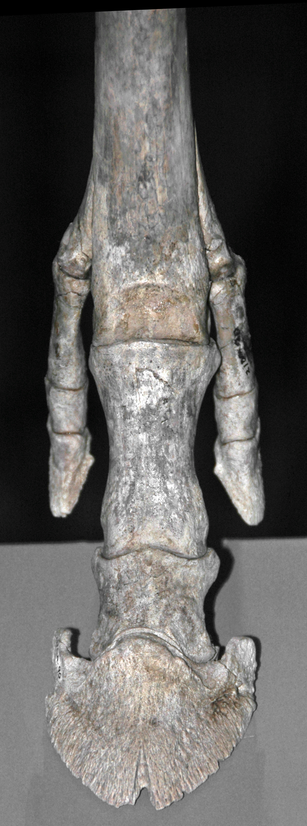
_foot_bones_(Ash_Hollow_Formation,_Miocene,_11.83_Ma;_Ashfall_Fossil_Beds,_Nebraska,_USA).jpg)
Atavisms have been recognized since antiquity, though they were interpreted in widely different ways before the advent of evolutionary theory. Pre-Darwinian naturalists sometimes described them as “reversions” or sports of nature, oddities without systematic meaning. Charles Darwin devoted considerable attention to reversion in On the Origin of Species and The Variation of Animals and Plants under Domestication, arguing that the persistence of dormant characters provided evidence of common ancestry. Modern developmental genetics has vindicated his interpretation in remarkable molecular detail, revealing the specific genes and regulatory networks responsible for the ancestral traits that occasionally resurface.
Definition and mechanism
A genuine atavism must meet two criteria. First, the trait must have been present in the organism’s actual evolutionary ancestors—confirmed by fossil or comparative evidence—and must have been absent from the lineage for a substantial period. Second, the reappearance must result from the reactivation of existing ancestral gene networks, not from the independent origin of a superficially similar trait. This distinction matters because it separates atavisms from convergent evolution and from pathological novelties that merely resemble ancestral states.12
The molecular mechanism underlying most atavisms involves changes in gene regulation rather than changes in the protein-coding genes themselves. In the course of evolution, a trait can be lost by several routes: the relevant structural genes may accumulate disabling mutations, becoming pseudogenes; or the regulatory elements that switch those genes on during development may be modified so the genes are no longer activated in the appropriate tissue at the appropriate time. Atavisms most commonly arise from the second route. If the structural genes remain largely intact but are normally kept silent by regulatory repressors, a disruption of those repressors—by a regulatory mutation, an environmental perturbation, or an experimental intervention—can restore gene expression and resurrect the ancestral structure.13, 14 The structural genes persist because they are often pleiotropic, serving other developmental functions in which their protein products are still needed, even after the ancestral structure was lost.
This framework explains both why atavisms occur and why they are rare. The ancestral developmental network must be preserved well enough to produce a recognizable structure when reactivated, but the regulatory silencing must be stable enough that reactivation is an infrequent accident rather than a routine developmental outcome. The result is a latent capacity for ancestral trait production that surfaces occasionally as a developmental anomaly.
Classic examples
Human coccygeal projections. The human coccyx is a fused series of three to five vestigial vertebrae representing the remnant of the tail present in the primate ancestors of Homo sapiens. In most humans the coccyx is entirely internal and bears no resemblance to a functional tail. In rare cases, however, infants are born with a soft tissue projection extending beyond the coccyx that contains muscle, connective tissue, blood vessels, and sometimes additional vertebrae beyond the normal coccygeal series.4 These structures are not simple skin tags or cysts; they are histologically organized projections that can be moved voluntarily by the individual in some documented cases, indicating that the musculature associated with ancestral tail movement has also been partially reactivated. The existence of these projections would be inexplicable if humans had been independently created without a common ancestor with tailed primates, but they follow directly from a model in which tail development genes have been suppressed in the lineage but not deleted.4, 3
Hind limbs in cetaceans. Whales and dolphins descended from terrestrial quadruped ancestors approximately 50 million years ago, a transition documented in exceptional detail by the fossil record of cetaceans.6, 15 During the course of this transition, the hind limbs were progressively reduced and eventually lost from the body wall, leaving only small internal pelvic remnants in modern species. However, occasional individual dolphins and whales are found with externally visible hind limb projections containing recognizable skeletal elements—femur, tibia, and in some cases even digit-like structures.5 These structures are atavisms: the reactivation of a developmental program that built functional hind limbs in the ancestors of whales. Their presence demonstrates that the genetic instructions for hind limb development have been maintained in the cetacean genome for tens of millions of years after hind limbs ceased to be part of the normal adult phenotype.5
Extra toes in horses. The modern horse (Equus caballus) is a single-hoofed animal, walking on the tip of one digit—the middle toe of what was ancestrally a five-toed foot. The horse lineage’s reduction from five toes to one over roughly 55 million years is one of the most thoroughly documented sequences in the fossil record, progressing through three-toed forms such as Merychippus and Pliohippus before reaching the single-toed condition in Equus.16 Modern horses, however, occasionally develop supernumerary toes—additional fully formed digits lateral to the main hoof, sometimes complete with their own hoof capsule, fetlock joint, and skeletal elements.7 These extra toes correspond precisely to the reduced lateral digits visible in fossil three-toed horses, confirming that their developmental genes have persisted and can be reactivated by regulatory changes. Julius Caesar reportedly possessed a horse with multiple hooves, an individual that historical sources treat as a portent; the modern developmental biology of equid polydactyly makes it unremarkable as a biological phenomenon.7
Hind limbs in snakes. All snakes descended from limbed lizard ancestors, and boid snakes such as pythons and boas retain small internal pelvic vestiges known as pelvic spurs, which are visible externally as claw-like protrusions flanking the cloaca. These spurs are the remnant of the ancestral pelvic and hind limb skeleton. Developmental studies of python embryos have shown that hind limb buds form normally in early development, with correct Hox gene expression along the proximal–distal axis, but that growth of the limb bud arrests at an early stage due to absence of the signaling molecule Sonic hedgehog (Shh) in the zone of polarizing activity.8 The ancestral limb-building gene network is therefore still active in snakes—it simply receives an incomplete set of growth signals. This explains why snake embryos briefly grow hind limb buds at all, and it provides a precise molecular account of how the transition from limbed ancestor to limbless descendant occurred through regulatory rather than structural gene changes.8, 10
Experimental induction of atavisms
One of the most striking confirmations of the atavism model came from experiments that did not wait for nature to produce a reversion but instead induced one deliberately in the laboratory. In 1980, Edward Kollar and Clifford Fisher reported a landmark experiment in which embryonic chicken tissue was combined with mouse dental mesenchyme in tissue recombination assays.1 Birds are toothless, having lost functional teeth roughly 80 million years ago; their beaks are keratinized structures with no enamel or dentine. Yet when chicken oral epithelium was placed in contact with mouse dental papilla cells—the mesenchymal cells that instruct overlying epithelium to form teeth—the chicken epithelium responded by producing structures with enamel, dentine, and crown morphology recognizable as teeth. The mouse mesenchyme had provided the inductive signals, but the chicken epithelium had executed the response using its own developmental genes, which had lain dormant for 80 million years of avian evolution.
A subsequent and methodologically cleaner set of experiments by Harris and colleagues in 2006 demonstrated that chicken teeth could be induced without the introduction of any foreign tissue at all.2 By manipulating signaling pathways active in the developing beak of the chicken embryo—specifically, activating the pathway associated with ectodermal organogenesis—they induced the formation of tooth-like structures in the beaks of mutant embryos carrying a recessive mutation called talpid2. The induced structures displayed enamel knots, the signaling centers that pattern mammalian tooth cusps, and produced mineralized tissue organized as genuine teeth with crowns of the type seen in Cretaceous toothed birds such as Hesperornis. The chicken genome had not merely retained the structural genes for tooth formation; it had retained a substantially complete tooth development program that could be unlocked by altering the regulatory inputs that normally suppress it.2
Parallel experiments in other systems have produced similarly striking results. Researchers working with threespine sticklebacks (Gasterosteus aculeatus) demonstrated that the loss of pelvic structures in freshwater populations of this fish—a loss that occurred independently many times in different lake populations over the past 10,000 years—results from changes in a regulatory enhancer that normally activates the gene Pitx1 in the pelvic field. When this regulatory change was reversed experimentally, pelvic structures were restored.11 The repeated, parallel loss of pelvis in independent freshwater stickleback populations, each targeting the same regulatory element, illustrates how ancestral developmental programs can be silenced and reactivated by changes in a single regulatory switch, and how the underlying structural genes persist intact through that process.
Molecular basis: Hox genes and conserved networks
The molecular architecture that makes atavisms possible rests on a set of deeply conserved developmental gene networks, the most prominent of which are the Hox genes. Hox genes encode transcription factors that specify positional identity along the major body axes in almost all animals, from insects to humans. They are organized in chromosomal clusters whose spatial arrangement in the genome corresponds to the sequence of body regions they pattern—a property called colinearity. This organization is conserved across 600 million years of animal evolution, indicating that Hox gene networks are subject to strong constraints that prevent their disruption.9
The stability of Hox networks across geological time is precisely what enables atavisms. The same Hox gene network that built tetrapod forelimbs in the common ancestor of snakes and lizards is still present in the snake genome; it has not been corrupted or deleted by 150 million years of limbless snake evolution. The same Hox network that built functional hind limbs in the terrestrial ancestors of whales persists in the dolphin genome. The same tooth-patterning network that produced the dentition of toothed Cretaceous birds persists in the chicken genome. These ancestral networks are conserved because their gene products serve other developmental roles, and because the regulatory machinery that suppresses them in their ancestral context is itself a modular addition that can be disrupted without dismantling the network itself.9, 10
The distinction between cis-regulatory and protein-coding evolution is central to understanding atavisms at the molecular level. When a trait is lost by accumulation of disabling mutations in the protein-coding sequences of the relevant structural genes—as has occurred for many olfactory receptor genes in primates or many taste receptor genes in various lineages—the result is a pseudogene whose product is non-functional. Reactivating such a gene could not restore an ancestral trait because the protein it encodes no longer works. Atavisms, by contrast, arise when the trait was lost by cis-regulatory changes that silenced otherwise intact genes. In these cases the protein-coding sequences remain functional, and reactivation of gene expression can resurrect the ancestral developmental pathway in working order.13, 14 The experimental induction of chicken teeth and stickleback pelvis are possible precisely because the structural genes in question were silenced by regulatory modifications rather than corrupted by coding mutations.
Atavisms and vestigial structures
Atavisms are conceptually related to, but distinct from, vestigial structures. A vestigial structure is a morphological feature present in all or most members of a population in reduced, simplified, or functionless form—the coccyx in all humans, the pelvic spurs present in all pythons, the hind limb remnants present internally in all cetaceans. A vestigial structure is a species-level trait, normal for the population even if functionless or reduced compared to the homologous structure in ancestor species. An atavism, by contrast, is an individual-level anomaly: the appearance, in a particular individual, of a more fully developed version of the ancestral structure that is normally reduced or absent at the population level.12
The distinction is one of developmental degree rather than kind. The coccyx is a vestigial tail in all humans; a coccygeal projection with additional vertebrae and musculature in a rare individual is an atavism of that same ancestral structure. Internal pelvic remnants present in all cetaceans are vestigial hind limbs; an externally protruding hind limb bud in an individual dolphin is an atavism. In both cases the underlying explanation is the same: retained ancestral developmental genes that no longer produce their full ancestral phenotype under normal developmental conditions but that retain the capacity to do so under altered regulatory conditions. Vestigial structures and atavisms are thus two manifestations of the same phenomenon—the persistence of ancestral genetic information across evolutionary time—differing only in whether the residual expression of that information is universal and reduced (vestigial) or individual and restored (atavistic).12, 13
This relationship means that the study of atavisms and vestigial structures is mutually reinforcing. The existence of a vestigial structure provides the prediction that an atavistic variant might be induced; the experimental induction of an atavism confirms that the developmental program underlying the vestigial structure is still encoded in the genome. The Kollar-Fisher chicken tooth experiments, for instance, are directly interpretable as experimental atavism—the experimental restoration of a structure that persists vestigially only in early chicken embryos—and they confirm that the avian tooth-forming program exists in a dormant, suppressible state rather than having been irreversibly dismantled.
Atavisms as evidence for common descent
The evidential weight of atavisms derives from a fundamental logical asymmetry: atavisms are straightforwardly expected under common descent but have no coherent explanation under special creation or under models that deny evolutionary relationships among the organisms that share them. If whales were independently created without a common ancestor with terrestrial mammals, there would be no reason for their genomes to contain the complete developmental program for building tetrapod hind limbs—a program that occasionally reasserts itself in individual animals as a recognizable limb bud containing femur, tibia, and digit precursors. The program is present because whales descended from tetrapods; the occasional atavistic hind limb is a developmental reminder of that ancestry rendered in bone and cartilage.5, 15
The convergence between atavistic morphology and fossil morphology provides an independent cross-check. The extra toes that appear atavistically in modern horses correspond in their position and skeletal organization to the lateral digits preserved in fossil three-toed and four-toed horses. The rudimentary hind limb elements that appear atavistically in cetaceans correspond to the reduced but present hind limb skeleton of early fossil whales such as Basilosaurus.15 The tooth morphology of experimentally induced chicken teeth corresponds to the crown patterns documented in fossils of toothed Cretaceous birds.2 In each case, the atavistic trait independently recovers the ancestral morphology documented in the fossil record, without any knowledge of that record being built into the developmental mechanism. This convergence between developmental genetics and palaeontology, achieved through entirely independent lines of evidence, provides strong corroboration that the atavistic traits genuinely reflect ancestral states rather than coincidental developmental anomalies.1, 13
Atavisms also constrain hypotheses about the mechanisms of evolutionary change. They demonstrate that major morphological transitions—loss of hind limbs in cetaceans and snakes, loss of teeth in birds, reduction from five to one toe in horses—can be accomplished by regulatory modifications without destroying the underlying gene networks. This finding aligns with a broader body of evidence from evolutionary developmental biology indicating that morphological evolution often proceeds through changes in the timing, location, and level of gene expression rather than through changes in the structure of the proteins those genes encode. The gene networks responsible for building ancestral structures are ancient, shared, and surprisingly robust—and atavisms are the most dramatic demonstration of just how robust they are.8, 9, 11
The fact that atavisms can be induced experimentally adds a further dimension to their evidential value. The experimental induction of chicken teeth or stickleback pelvic structures is not merely an observation of nature; it is a controlled test of the hypothesis that ancestral gene networks persist in suppressed form. The experiments predict specific outcomes—tooth formation in a normally toothless bird, pelvic growth in a normally pelvic-reduced fish—and those predictions are confirmed. This predictive and testable character is the hallmark of scientific evidence, and it distinguishes atavisms from mere anecdote. The genome, interpreted through the lens of evo-devo, functions as a molecular archive of evolutionary history; atavisms are the moments when that archive’s contents briefly become legible in the phenotype of a living animal.1, 2, 11
References
Induction of tooth morphogenesis in mouse epithelium by experimentally induced dental papilla
Repeated morphological evolution through cis-regulatory changes in a pleiotropic gene
Evolutionary history of the horse: new perspectives on the origins of equid diversity