Overview
- The cell is the fundamental structural and functional unit of all known life, a principle formalized by Schleiden, Schwann, and Virchow in the nineteenth century and since confirmed by every branch of biology — no entity smaller than a cell is independently alive, and no organism exists that is not composed of one or more cells.
- Life on Earth is divided between two profoundly different cellular architectures: prokaryotic cells (bacteria and archaea), which lack a membrane-bound nucleus, and eukaryotic cells, which compartmentalize their DNA within a nucleus and contain organelles such as mitochondria and chloroplasts that originated through endosymbiosis.
- Because all heritable variation, natural selection, and replication occur at the cellular level or are organized by cellular machinery, the cell is the irreducible unit upon which evolution acts — every adaptation, every lineage, and every major transition in the history of life is ultimately a story written in cells.
The cell is the smallest unit of matter that can be called alive. Every organism on Earth, from the simplest bacterium to the largest whale, is composed of one or more cells, and no living process occurs outside of one.9 This principle, codified in the nineteenth century as cell theory, stands as one of the most fundamental and universally confirmed generalizations in all of biology. The cell is where metabolism happens, where genetic information is stored and copied, where proteins are synthesized, and where evolution ultimately plays out. Understanding the cell is therefore prerequisite to understanding life itself.
.jpg)
Cell theory
The recognition that all living things are composed of cells emerged gradually over two centuries of microscopy. Robert Hooke first used the word "cell" in 1665 to describe the boxlike compartments he observed in thin slices of cork under his microscope, though what he actually saw were the empty cell walls of dead plant tissue.9 Antonie van Leeuwenhoek, working in the 1670s with single-lens microscopes of his own design, was the first to observe living single-celled organisms, which he called "animalcules," in pond water, saliva, and other fluids. These observations established that microscopic life existed in extraordinary abundance, but it was not yet understood that cells were a universal structural principle.
That generalization came in 1838 and 1839, when the botanist Matthias Schleiden and the zoologist Theodor Schwann independently proposed that cells are the fundamental units of both plant and animal life.1, 2 Schleiden concluded from his studies of plant tissues that every structural element of a plant is composed of cells or their products.1 Schwann extended this principle to the animal kingdom, arguing that the same basic unit of organization underlies all living tissue, whether leaf or liver, and that the differences between organisms are differences in how cells are arranged and specialized, not differences in fundamental architecture.2 Together, these two propositions formed the first and second tenets of classical cell theory: all living things are composed of cells, and the cell is the basic unit of life.
The third tenet was added in 1855 by the physician Rudolf Virchow, who declared omnis cellula e cellula — every cell arises from a pre-existing cell.3 This statement was a direct repudiation of the then-popular doctrine of spontaneous generation, which held that living organisms could arise de novo from non-living matter. Virchow's insight reframed the cell not merely as a structural element but as a unit of biological continuity: cells beget cells, and the unbroken chain of cellular descent stretches back to the origin of life itself.
Taken together, the three tenets of cell theory — all life is cellular, the cell is the fundamental unit, and all cells come from cells — remain foundational to modern biology, refined but never overturned by a century and a half of subsequent research.9 Modern cell biology has added nuance: viruses, for example, are not cellular and cannot reproduce independently, yet they require host cells to replicate and are composed of molecular machinery that ultimately derives from cellular life.9 Prions and viroids further complicate the boundary between living and non-living matter. None of these entities, however, challenge the central claim of cell theory. They are parasites of cellular systems, not alternatives to them, and their existence reinforces rather than undermines the principle that the cell is the irreducible unit of independent life.
Prokaryotic cells
The simplest and most ancient cellular architecture is the prokaryotic cell, a design shared by bacteria and archaea. Prokaryotic cells lack a membrane-bound nucleus; their DNA resides in a region of the cytoplasm called the nucleoid, freely accessible to the cell's transcriptional and translational machinery.9 They are typically small, ranging from about 0.2 to 10 micrometres in diameter, and they lack the elaborate internal membrane systems that characterize eukaryotic cells.14 Despite this apparent simplicity, prokaryotes are extraordinarily successful. They colonize virtually every habitat on Earth, from deep-sea hydrothermal vents to Antarctic ice, from the human gut to the upper atmosphere, and they outnumber all eukaryotic cells on the planet by orders of magnitude.9
For most of the twentieth century, all prokaryotes were classified together as bacteria. This changed dramatically in 1977, when Carl Woese and George Fox used ribosomal RNA sequence comparisons to demonstrate that what had been called "bacteria" actually comprised two fundamentally distinct domains of life: the Bacteria and the Archaea.4 Although archaea superficially resemble bacteria in cell size and the absence of a nucleus, their molecular biology is in many respects more similar to that of eukaryotes. Archaeal ribosomes, DNA replication machinery, and transcription apparatus share key features with their eukaryotic counterparts that are absent in bacteria.5 This molecular kinship between archaea and eukaryotes would later prove central to understanding the origin of the eukaryotic cell. Woese formalized the three-domain system in 1990, proposing that all cellular life be classified into the domains Bacteria, Archaea, and Eucarya, a framework that remains the standard today.5
Prokaryotic cells reproduce by binary fission, a process in which the circular chromosome is replicated and the cell divides into two genetically identical daughter cells. Binary fission is rapid — Escherichia coli can divide every 20 minutes under optimal laboratory conditions — and does not involve the complex chromosome-condensation and spindle-assembly machinery of eukaryotic mitosis.9, 14 This speed of reproduction, combined with large population sizes and the capacity for horizontal gene transfer, gives prokaryotic populations an enormous capacity for rapid evolutionary adaptation, as vividly demonstrated by the evolution of antibiotic resistance in clinical settings.9
Eukaryotic cells
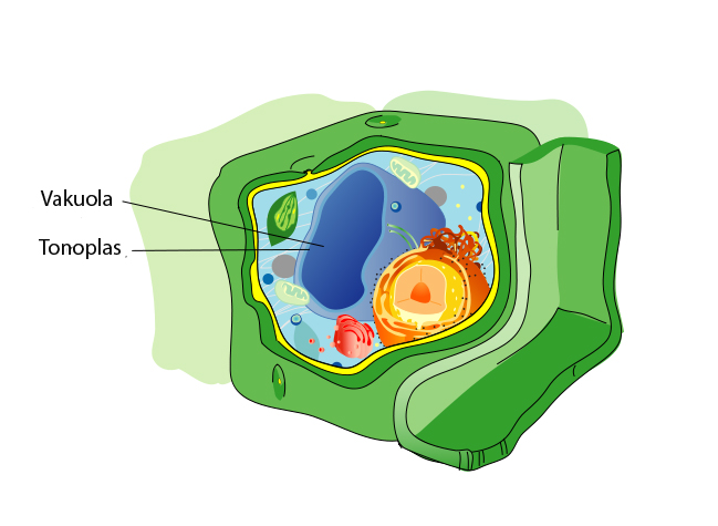
Eukaryotic cells are distinguished from prokaryotes by a defining feature: a membrane-bound nucleus that encloses the cell's chromosomal DNA, physically separating transcription from translation.9 But the nucleus is only the most conspicuous of many internal membrane-bound compartments. Eukaryotic cells contain an elaborate endomembrane system that includes the endoplasmic reticulum, the Golgi apparatus, lysosomes, and peroxisomes, each performing specialized biochemical functions within its own enclosed environment. This internal compartmentalization allows eukaryotic cells to run incompatible chemical reactions simultaneously in separate compartments, a capacity that prokaryotes, with their single undivided cytoplasm, generally lack.9
The most consequential organelles in the eukaryotic cell are the mitochondria. Present in virtually all eukaryotes, mitochondria are the sites of oxidative phosphorylation, the metabolic process that generates the majority of a cell's ATP. As described in detail in the article on endosymbiosis, mitochondria originated as free-living alphaproteobacteria that were engulfed by an ancestral host cell and progressively integrated into its biology.7, 11 They retain their own small circular genome, their own 70S ribosomes, and they divide by binary fission independently of the host cell's nuclear division, all vestiges of their bacterial ancestry. Photosynthetic eukaryotes additionally contain chloroplasts, which descended from a cyanobacterial endosymbiont and carry out oxygenic photosynthesis.7
Eukaryotic cells are typically 10 to 100 micrometres in diameter, roughly ten times larger than most prokaryotes in linear dimension and a thousand times greater in volume.14 This increase in size was made possible, at least in part, by the energetic contribution of mitochondria. A eukaryotic cell with thousands of mitochondria can sustain a far larger genome and a far more complex proteome than a prokaryotic cell that must generate all its ATP across a single plasma membrane.11 The eukaryotic cytoskeleton, a dynamic network of actin filaments, microtubules, and intermediate filaments, provides the structural framework for this larger cell, enabling it to maintain its shape, transport cargo internally, and divide its contents with precision during cell division.9
The cell membrane
Every cell, whether prokaryotic or eukaryotic, is bounded by a plasma membrane that separates the interior of the cell from its external environment. This membrane is not a rigid wall but a dynamic, fluid structure composed primarily of a lipid bilayer — two layers of phospholipid molecules arranged with their hydrophobic fatty acid tails facing inward and their hydrophilic head groups facing outward toward the aqueous environments on either side.9 The spontaneous self-assembly of lipid bilayers in water is driven by the hydrophobic effect and requires no biological machinery, a property that has significant implications for understanding how the earliest protocells may have formed during the origin of life.13
The modern understanding of membrane architecture is based on the fluid mosaic model, proposed by S. Jonathan Singer and Garth Nicolson in 1972.6 In this model, the lipid bilayer is a two-dimensional fluid in which individual lipid molecules can move laterally within their own leaflet, and proteins are embedded in or associated with the bilayer in a mosaic pattern. Integral membrane proteins span the bilayer entirely and serve as channels, transporters, receptors, and enzymes, while peripheral membrane proteins associate with the membrane surface and participate in signaling and structural roles.6, 9 Although the fluid mosaic model has been refined considerably since 1972 — modern views emphasize the existence of lipid rafts, membrane domains of varying fluidity, and a far greater density of membrane proteins than originally depicted — its core insight that the membrane is a dynamic, heterogeneous fluid rather than a static barrier remains the accepted framework.6
One of the most revealing differences between the three domains of life is the chemical composition of their membrane lipids. Bacterial and eukaryotic membranes are built from fatty acid ester lipids, in which fatty acids are linked to a glycerol backbone by ester bonds. Archaeal membranes, by contrast, are built from isoprenoid ether lipids, in which branched isoprenoid chains are linked to glycerol by ether bonds, and the stereochemistry of the glycerol backbone is inverted.5, 9 These differences in membrane chemistry are so fundamental that they have been interpreted as evidence that the last universal common ancestor (LUCA) may have had a heterogeneous or leaky membrane, with the modern membrane types emerging independently in the bacterial and archaeal lineages after their divergence.15
Cell division
The continuity of life depends on the ability of cells to divide, producing new cells that carry a faithful copy of the parental genetic information. The mechanisms of cell division differ substantially between prokaryotes and eukaryotes, reflecting their different levels of genomic and structural complexity.
Prokaryotic cells divide by binary fission, a relatively straightforward process. The single circular chromosome is replicated, the two copies are segregated to opposite ends of the cell, and a septum forms at the midpoint, cleaving the cell into two daughter cells. The protein FtsZ, a homologue of the eukaryotic cytoskeletal protein tubulin, assembles into a contractile ring at the division site and plays a central role in coordinating septum formation.9, 10 The entire process can be completed in as little as 20 minutes in fast-growing bacteria, and the fidelity of chromosome segregation is ensured by active mechanisms that are simpler than, but functionally analogous to, the mitotic spindle of eukaryotes.14
Eukaryotic cells face a far more complex task. Their genomes are distributed across multiple linear chromosomes, each of which must be precisely replicated and segregated. Mitosis, the form of cell division that produces two genetically identical daughter cells, accomplishes this through a carefully orchestrated sequence of chromosome condensation, spindle assembly, chromosome alignment at the metaphase plate, sister chromatid separation, and cytokinesis.9 The mitotic spindle, a bipolar structure made of microtubules, physically attaches to each chromosome at a specialized protein complex called the kinetochore and pulls sister chromatids to opposite poles of the cell. Checkpoint mechanisms at multiple stages of mitosis ensure that division does not proceed until every chromosome is properly attached, a safeguard whose failure can lead to aneuploidy and, in multicellular organisms, cancer.9
Meiosis, a specialized form of cell division unique to sexually reproducing eukaryotes, produces four genetically distinct haploid cells from a single diploid precursor. It achieves this through two consecutive rounds of chromosome segregation: the first separates homologous chromosomes (reducing the chromosome number by half), and the second separates sister chromatids (as in mitosis).9 Critically, meiosis incorporates recombination — the physical exchange of DNA segments between homologous chromosomes during prophase I — which shuffles allelic combinations and generates the heritable genetic variation upon which natural selection acts. The evolution of meiotic sex was thus a pivotal innovation in the history of life, vastly accelerating the rate at which beneficial mutations could be combined and deleterious ones purged from populations.12
The endosymbiotic bridge
The most profound divide in cellular biology is between prokaryotes and eukaryotes. Eukaryotic cells possess a suite of features — a nucleus, an endomembrane system, a dynamic cytoskeleton, mitochondria — that are entirely absent from prokaryotes, and no living intermediate forms exist. How did this enormous gap arise? The answer, supported by overwhelming molecular and phylogenetic evidence, is endosymbiosis: the eukaryotic cell is a chimera, forged by the merger of an archaeal host cell and one or more bacterial endosymbionts.7, 11
The host cell in this partnership was an archaeon. Phylogenomic analyses consistently place eukaryotes within or as the sister group of the Asgard archaea, a superphylum whose genomes encode homologues of many proteins previously thought to be exclusive to eukaryotes, including components of the membrane-trafficking system, the ubiquitin pathway, and actin-related cytoskeletal elements.8 The endosymbiont was an alphaproteobacterium that became the mitochondrion. This event was singular and unrepeated: every known eukaryote either possesses mitochondria or possesses organelles of clear mitochondrial descent that have been secondarily reduced, indicating that the last eukaryotic common ancestor already harboured a mitochondrial endosymbiont.11
The acquisition of the mitochondrial endosymbiont was transformative. Aerobic respiration via the mitochondrial electron transport chain generates roughly 15 times more ATP per molecule of glucose than fermentation alone, providing eukaryotic cells with an energy budget vastly larger than that available to any prokaryote of comparable size.11, 9 This energetic surplus is widely hypothesized to have been a prerequisite for the evolution of large cell size, genomic complexity, and the elaborate internal membrane systems that distinguish eukaryotes from prokaryotes.11 A second endosymbiotic event, in which an ancestor of modern cyanobacteria was engulfed by a eukaryotic host that already possessed mitochondria, gave rise to the chloroplast and enabled eukaryotes to perform oxygenic photosynthesis, ultimately establishing the plant and algal lineages that sustain most of Earth's ecosystems.7
The endosymbiotic origin of mitochondria and chloroplasts means that every eukaryotic cell is, in a real sense, a community of formerly independent organisms. The nuclear genome is predominantly archaeal in its information-processing genes, while many metabolic genes trace their ancestry to the bacterial endosymbionts.11, 15 This chimeric nature — archaeal in heritage, bacterial in metabolic capacity — is one of the defining features of the eukaryotic cell and a vivid illustration of how evolution can build fundamentally new kinds of complexity not through gradual modification alone but through the wholesale merger of distinct lineages.
Prokaryotic vs. eukaryotic cells9, 5, 14
| Feature | Prokaryotes (Bacteria & Archaea) | Eukaryotes |
|---|---|---|
| Nucleus | Absent; DNA in nucleoid region | Present; membrane-bound |
| Typical cell diameter | 0.2–10 µm | 10–100 µm |
| Genome structure | Single circular chromosome (usually) | Multiple linear chromosomes |
| Membrane-bound organelles | Absent | Present (mitochondria, ER, Golgi, etc.) |
| Ribosomes | 70S | 80S (cytoplasmic); 70S (mitochondrial) |
| Cell division | Binary fission | Mitosis / meiosis |
| Membrane lipids | Ester-linked (Bacteria); ether-linked (Archaea) | Ester-linked |
| Cytoskeleton | Minimal (FtsZ, MreB) | Complex (actin, tubulin, intermediate filaments) |
Cells as the unit of evolution
The cell is not merely a structural unit; it is the unit upon which evolution operates. Natural selection acts on organisms, and every organism is built from cells. The heritable variation on which selection depends — mutations, recombinations, epigenetic modifications — arises during the processes of DNA replication and cell division.9, 12 When a mutation confers a fitness advantage, it spreads through a population because the cells carrying it outcompete those that do not, whether that population is a colony of bacteria doubling every hour or a species of mammal reproducing over decades. In this sense, every adaptation in the history of life, from antibiotic resistance in Staphylococcus to the evolution of the eye in vertebrates, is ultimately a story about cells that divided more successfully than their competitors.
The major transitions in evolution, as catalogued by John Maynard Smith and Eörs Szathmáry, are almost all transitions in how cells relate to one another.12 The origin of the first cell was the origin of life itself. The merger of archaeal and bacterial cells through endosymbiosis created the eukaryotic cell. The aggregation of eukaryotic cells into cooperative collectives gave rise to multicellularity. The differentiation of cells within multicellular organisms produced tissues and organs. At each stage, the cell remained the indivisible protagonist: the entity that replicates, the entity that mutates, the entity that is selected.
Even in multicellular organisms, where selection is often described as acting on the whole organism, the cellular perspective remains essential. Cancer, for example, is fundamentally a disease of cellular evolution: individual cells within a multicellular body acquire mutations that cause them to proliferate without restraint, effectively reverting to a program of unicellular natural selection at the expense of the organism.9 The tension between cooperation and competition among cells within a multicellular body is one of the central problems in evolutionary biology, and its resolution — through mechanisms such as programmed cell death, immune surveillance, and the germ-soma distinction — was essential for the emergence of complex multicellular life.12
From the first protocells that enclosed self-replicating molecules in a lipid membrane to the trillions of specialized cells that compose a human body, the cell has been the continuous thread of biological existence. It is the arena in which the chemistry of the RNA world became biology, the vessel through which genetic information has been transmitted across nearly four billion years, and the fundamental unit from which every living structure on Earth is assembled. Understanding the cell is, in the deepest sense, understanding what it means to be alive.
References
Mikroskopische Untersuchungen über die Uebereinstimmung in der Struktur und dem Wachsthum der Thiere und Pflanzen
Die Cellularpathologie in ihrer Begründung auf physiologische und pathologische Gewebelehre
Towards a natural system of organisms: proposal for the domains Archaea, Bacteria, and Eucarya
The last universal common ancestor between ancient Earth chemistry and the onset of genetics